


中国医疗器械博览会|超声弹性成像:简史、临床价值与局限边界
2025-08-22
中国医疗器械博览会带您了解超声弹性成像:简史、临床价值与局限边界如下。
What it is and where it began.
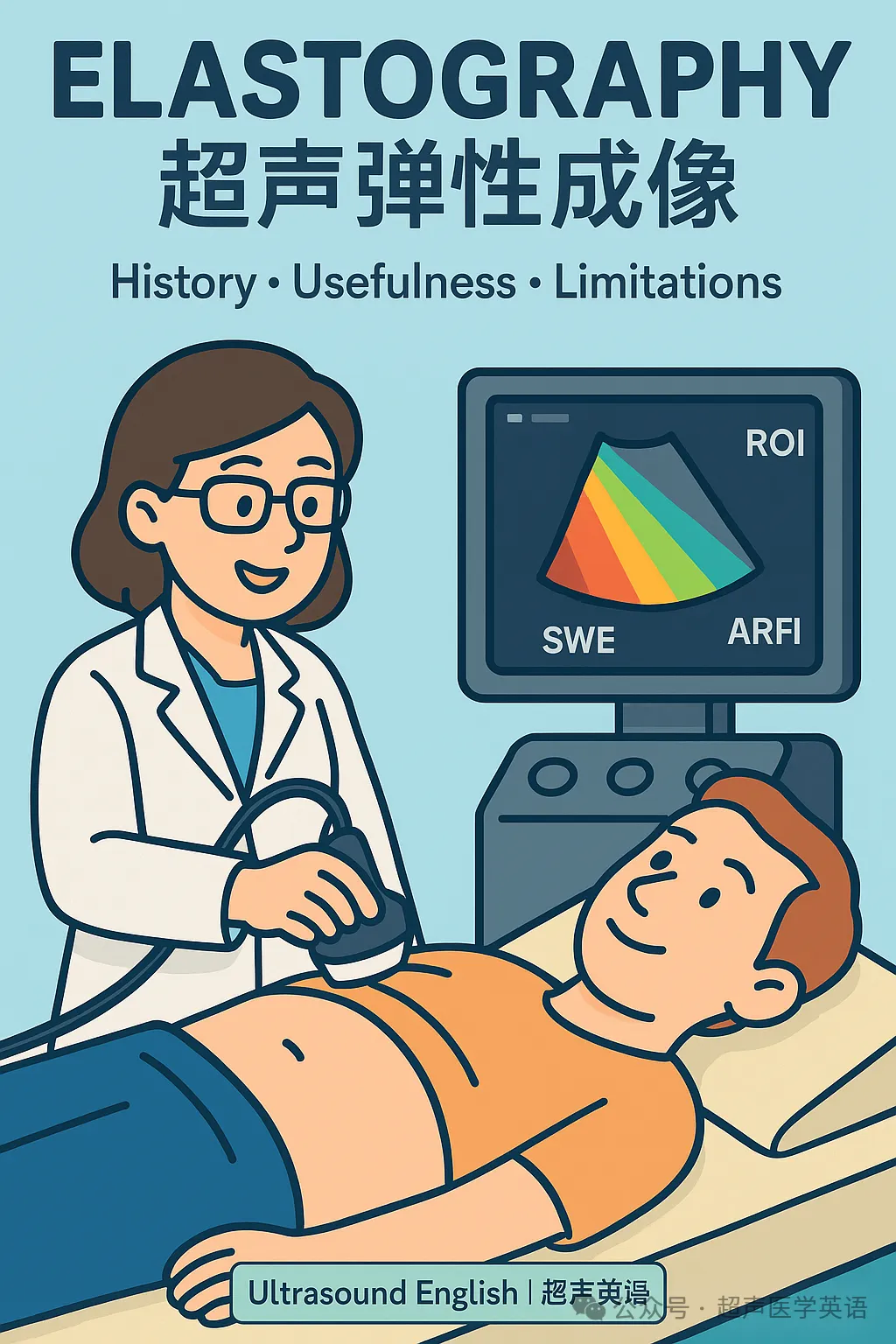
Elastography is an ultrasound technique that visualizes and quantifies tissue stiffness—an attribute tightly linked to pathology such as fibrosis, inflammation, and malignancy. The idea surfaced in the late 1980s and early 1990s: if a lesion feels “hard” under a surgeon’s fingers, could imaging capture that hardness objectively? Early strain elastography answered with qualitative color maps created by gentle compression from the probe or physiologic motion (heartbeat, respiration). These first-generation systems demonstrated feasibility but were operator-dependent and hard to standardize.
The quantitative turn.
In the 2000s, two milestones changed clinical adoption: acoustic radiation force impulse (ARFI) and shear wave elastography (SWE). ARFI used focused ultrasound pushes to generate tiny tissue displacements, while SWE measured the shear wave velocity (SWV) that propagates through tissue; faster waves indicate stiffer tissue. Because SWV can be converted to kilopascals via elastic models, stiffness became a number rather than a color, enabling reproducible thresholds, multi-center studies, and guidelines.
Why it matters.
The most transformative use is the non-invasive staging of liver fibrosis. Elastography reduces liver biopsy rates, helps screen for advanced fibrosis, and supports decisions about antiviral therapy and variceal risk. Beyond the liver, elastography adds value in breast (distinguishing benign from suspicious lesions), thyroid (risk stratification of nodules), prostate (target selection for biopsy), lymph nodes (reactive vs metastatic), and musculoskeletal applications (tendon, muscle, plantar fascia, and scar assessment). In oncology and interventional practice, stiffness mapping can guide biopsy, plan ablation margins, and monitor treatment response over time.
How to get reliable numbers.
Quality hinges on good technique. Keep probe pressure minimal to avoid artificial stiffness; align the region of interest (ROI) away from large vessels and lesion edges; mind depth, because very deep targets attenuate waves; ask for breath-hold during liver measurements; and use the system’s quality map/indicator to accept only robust acquisitions. Report the median of multiple measurements, note the IQR/median ratio (as a reliability metric), and always integrate findings with B-mode and Doppler.
What can go wrong.
Elastography has limitations. Results vary with vendor algorithms and presets; inter- and intra-observer variability remains an issue without training. Obesity, ascites, rib shadowing, or poor acoustic windows degrade shear waves. Precompression, anisotropy in tendons, and boundary effects near cysts or calcifications create artifacts. Stiffness is not diagnosis: inflammation, congestion, cholestasis, and post-prandial states can mimic fibrosis. For thyroid and breast, overlap exists between benign and malignant ranges, so elastography should complement, not replace cytology, histology, and risk scores.
Current best practices.
Use validated cut-off values for specific organs and machines; document machine model, probe, depth, patient position, and whether the patient was fasting for liver exams. When following patients, try to keep the same vendor and preset. For research or multicenter audits, build prospective registries capturing stiffness, clinical context, and outcomes.
Where it’s going.
Three trends stand out. First, multiparametric ultrasound—combining B-mode, Doppler, CEUS, and elastography—improves diagnostic confidence and procedural planning. Second, 3D/4D SWE and fusion imaging (US-CT/MR) offer volumetric stiffness maps and precise lesion registration for focal therapy. Third, AI assistance is emerging: automated ROI placement, artifact detection, and trajectory planning for needle paths could reduce variability and shorten learning curves. As standards harmonize and reference databases grow, elastography will move from “helpful add-on” to a routine quantitative vital sign for tissue.
Bottom line.
Elastography converts a clinician’s fingertip impression into numbers you can track. When performed and interpreted well, it speeds decisions, reduces unnecessary biopsies, and monitors disease and therapy with minimal burden to patients. Use it thoughtfully—respecting artifacts, physiology, and clinical context—and it will earn a reliable seat in your day-to-day ultrasound toolkit.
Elastography Vocabulary
-
Elastography /ɪˌlæˈstɒɡrəfi/ 弹性成像(elastic+-graphy)
-
Strain /streɪn/ 应变(拉伸)
-
Strain elastography /streɪn ɪˌlæˈstɒɡrəfi/ 应变弹性成像
-
Shear wave /ʃɪər weɪv/ 剪切波
-
Shear wave elastography (SWE) /…/ 剪切波弹性成像
-
Acoustic radiation force impulse (ARFI) /…/ 声辐射力脉冲
-
Shear wave velocity (SWV) /…/ 剪切波速度
-
Elastic modulus /ɪˈlæstɪk ˈmɒdjʊləs/ 弹性模量
-
Young’s modulus /jʌŋz…/ 杨氏模量
-
Poisson’s ratio /ˈpwɑːsɒnz…/ 泊松比
-
Region of interest (ROI) /ˈriːdʒən…/ 感兴趣区
-
Quality map/indicator /ˈkwɒləti mæp/ 质量图/指标
-
Precompression /ˌpriːkəmˈprɛʃən/ 预压缩
-
Anisotropy /ˌænɪˈsɒtrəpi/ 各向异性
-
Attenuation /əˌtɛnjʊˈeɪʃən/ 衰减
-
Boundary effect /ˈbaʊndəri ɪˈfɛkt/ 边界效应
-
Aliasing /ˈeɪliəsɪŋ/ 混叠
-
Artifact /ˈɑːrtɪˌfækt/ 伪影
-
Repeatability /rɪˌpiːtəˈbɪləti/ 重复性
-
Reproducibility /ˌriːprəˌdjuːsəˈbɪləti/ 可重复性
-
Interobserver variability /ˌɪntərəbˈzɜːrvər…/ 检者间差异
-
Intraobserver variability /ˌɪntrəəbˈzɜːrvər…/ 检者内差异
-
Cut-off value /ˈkʌt ɒf ˈvæljuː/ 截止值
-
IQR/median ratio /aɪ kjuː ɑːr…/ 四分位距/中位数比
-
Non-invasive /nɒn ɪnˈveɪsɪv/ 无创的
-
Fibrosis staging /faɪˈbrəʊsɪs/ 纤维化分期
-
Cirrhosis /səˈroʊsɪs/ 肝硬化
-
Portal hypertension /ˈpɔːrtl…/ 门静脉高压
-
Acoustic window /əˈkuːstɪk…/ 声窗
-
Breath-hold /brɛθ hoʊld/ 屏气
-
Fasting state /ˈfæstɪŋ steɪt/ 空腹状态
-
Lymphadenopathy /ˌlɪmˌfædɪˈnɒpəθi/ 淋巴结病
-
Nodule /ˈnɒdʒuːl/ 结节
-
Benign vs malignant /bɪˈnaɪn… məˈlɪɡnənt/ 良性/恶性
-
Musculoskeletal (MSK) /ˌmʌskjʊləˈskɛlɪtəl/ 肌骨系统
-
Elastogram /ɪˈlæstəɡræm/ 弹性图
-
Multiparametric ultrasound /ˌmʌltipaːrəˈmɛtrɪk/ 多参数超声
-
Fusion imaging /ˈfjuːʒən/ 融合成像
-
Ablation planning /əˈbleɪʃən/ 消融规划
-
Treatment monitoring /ˈmɒnɪtərɪŋ/ 治疗监测
超声弹性成像:简史、临床价值与局限边界
进入21世纪,声辐射力脉冲(ARFI)与剪切波弹性成像(SWE)奠定了临床化的基础。ARFI通过聚焦超声“推一下”组织,SWE测量在组织中传播的剪切波速度;速度越快,组织越硬。借助弹性模型,速度可换算为千帕等单位,硬度从“颜色”变为“数字”,使阈值、研究与指南成为可能。
临床价值最突出的是肝纤维化的无创分期:减少活检、评估晚期纤维化与静脉曲张风险,并指导抗病毒治疗。除此之外,弹性成像在乳腺(良恶性鉴别)、甲状腺(结节分层)、前列腺(靶向活检选择)、淋巴结(反应性vs转移)、以及肌骨系统(肌腱与瘢痕评估)均有贡献。在肿瘤与介入场景中,硬度图有助于定位取材、规划消融边界并随访疗效。
获得可靠结果的关键是规范操作:保持最小探头压力,ROI避开大血管与病灶边缘;关注深度与声窗;肝脏测量要求屏气;仅接受系统显示高质量的波形;报告多次测量的中位数及IQR/中位数比。任何读数都必须结合B超与临床信息解读。
局限与陷阱:不同厂商算法与预设导致可比性受限;缺乏培训时检者间与检者内差异明显。肥胖、腹水、肋骨声影会降低剪切波质量;预压、各向异性、边界效应可造成假硬度。硬度并不等同诊断:炎症、充血、胆汁淤积、进食状态都可能“假装纤维化”。在甲状腺与乳腺,良恶性硬度仍有重叠,因此弹性成像应作为补充而非替代活检与其他成像。
发展趋势:其一,多参数超声将B超、血流、造影与弹性融合,提升诊断与手术规划;其二,3D/4D SWE与融合成像带来体积硬度图与高精度配准;其三,AI正用于自动ROI、伪影识别与穿刺路径规划,以降低变异、缩短学习曲线。随着标准统一与参考数据库扩大,弹性成像有望成为类似“生命体征”的组织硬度指标。
要点:只要尊重物理与生理、注意伪影并结合临床,弹性成像就能减少不必要的活检,加快决策,为患者带来更轻负担的诊断与随访。
图片来源:超声医学英语
<中国医疗器械博览会 >将于2025年9月24-26日在上海世博展览馆1&2号馆举办。现场汇聚近1000家来自全球近27个国家的优质品牌供应商,为中国医疗器械生产厂商提供产品研发、生产、注册所需的设计及软件服务、原材料、精密部件、自动化制造设备、超精加工技术、合同制造、测试和认证、政策法规和市场咨询服务,展品覆盖医疗器械设计与制造全产业链。预登记已开放,点击提前注册即可免费参观,立省百元门票费,现场注册需付费。
文章来源:超声医学英语
文章内容仅供知识交流分享使用,如涉侵权请联系我们删除。