


重磅!20232023上海医疗器械创新展速递新技术:一种用于梗塞心脏修复的具有ANGPTL4缓释的可涂抹和粘附的水凝胶心脏补片 | BAM
2023-11-20
近期,韩国光州科学技术院Jae Young Lee研究团队在科爱创办的期刊Bioactive Materials上发表文章: 心肌梗死再灌注后发生不可逆的病理重构,包括左心室扩张和心肌梗死后的过度炎症反应,常常导致致命性的功能损害。人们已广泛尝试利用心脏补片和抗炎药物输送来减轻心肌梗死后的病理性重构。在这项研究中,作者开发了一种使用葡聚糖醛(Dex-ald)和明胶的可涂性和粘附性水凝胶贴片,将抗炎蛋白ANGPTL4加入水凝胶中,直接向梗塞心脏持续释放,以减轻炎症。
01.研究内容简介
1. 引言
全球每年有超过1790万人死于心血管疾病。以心肌梗塞(MI)为代表的缺血性心脏病是心血管疾病相关死亡的主要原因。心肌梗死主要是由于冠状动脉内动脉粥样斑块破裂和血栓形成导致流向心肌的血流和氧气的阻塞。积极探索减轻梗死心脏的病理性重塑并促进心脏修复至关重要。特别是,一些心脏贴片已被应用于通过减少过度扩张来支持梗塞心脏的机械功能。然而,大多数粘附性水凝胶是使用化学交联剂和/或不可生物降解的合成聚合物生产的,这引起了人们对其生物相容性和实际应用的担忧。因此,开发一种生物相容性和粘附性的水凝胶心脏补片用于心脏修复仍然具有挑战性。
在这项研究中,作者设计了一种可涂抹的粘附性水凝胶,可以持续释放ANGPTL4,作为一种多功能心脏补片,用于心肌梗死后有效的心脏修复。具体地说,生物相容的天然聚合物(明胶和右旋糖醛(Dex-ald))被配制成自发形成凝胶,明胶/Dex-ald水凝胶和组织之间的非共价相互作用(如氢键和静电相互作用)和席夫碱的形成进一步允许凝胶与心脏组织的强大粘附性。他们研究的粘附性水凝胶心脏贴片(载ANGPTL4明胶/地塞米松)应该能有效地减轻病理性重构(如左室壁变薄和心腔扩张),并通过在心外膜持续释放抗炎因子ANGPTL4来抑制过度炎症,从而诱导心脏修复。
2. 结果和讨论
2.1. 可涂装水凝胶的设计与制备
他们使用明胶和葡聚糖作为基础水凝胶材料,然后进一步将右旋糖苷修饰为地塞米松,它可以通过席夫反应与明胶和心脏组织自发反应,分别与心外膜形成共价水凝胶网络和强烈的粘附键。这种混合后的自发反应使得水凝胶可以在左心外膜上涂抹(图1)。合适的原位凝胶反应动力学是获得可涂抹的水凝胶心脏补片的关键。当心外膜上的原位凝胶发生得太慢时,前体溶液可能在凝胶之前从心脏流出,导致心脏上的水凝胶不完整。另一方面,过快的凝胶化可能会导致水凝胶在心外膜上的不可控和不均匀的覆盖,以及对组织的粘附力不足。因此,他们试图通过考察前体浓度和相对分子质量等各种因素来获得最佳的凝胶化时间。首先,探讨了地塞米松和明胶组成对凝胶形成的影响。明胶中胺含量为0.26,乙醛含量为3.48 mmol/g(图S1)。在明胶含量恒定(10%)的情况下,dex-ald的浓度从1%到5%不等,与乙醛相比,胺基过多。混合物中较高的 dex-ald 含量导致弹性模量增加更快,凝胶化时间更短时间。此外,含有更多dex-ald的凝胶/ dex-ald水凝胶具有更高的模量(图2A和2B)。这些结果表明,醛密集存在于 dex-ald 链中的基团(0.58 个醛基团在葡萄糖单位)与胺基团的可及性和反应性有限在明胶中形成希夫碱键,需要更大的醛含量用于网络形成比胺基团。明胶的变化明胶的变化在 Dex-ALD 含量恒定的情况下,含量从2.5%到10% 不显著影响凝胶时间(图2C和2D)。然而,明胶/DEX-ALD的弹性模量随着明胶含量的增加而增加。
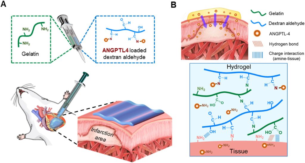
图1 Schematic representations of an adhesive paintable hydrogel. (A) Schematic illustration of a paintable hydrogel consisting of dextran-aldehyde and gelatin for the treatment of infarcted hearts. (B) Schematic illustration of sustained release of ANGPTL4 from a paintable hydrogel on epicardium to infarcted myocardium.
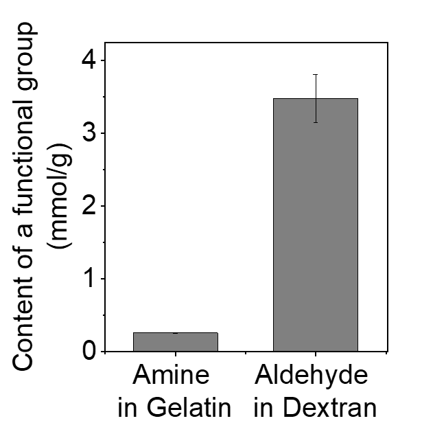
图S1 The content of a functional group (amine or aldehyde) in each polymer.
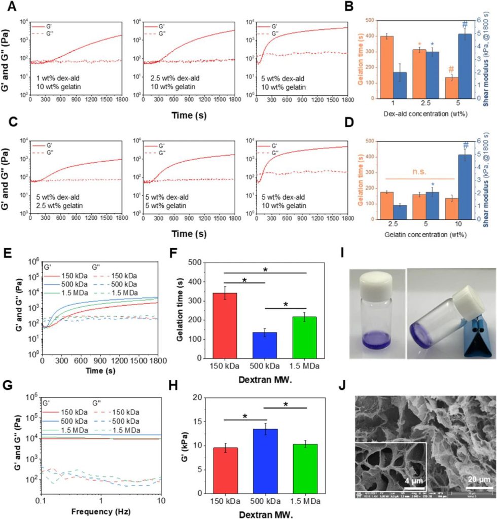
图2 Preparation and characterization of the paintable gelatin/dex-ald hydrogels. (A) Time-dependent shear moduli of various hydrogels prepared with 10% gelatin and different concentrations of dex-ald. G′ and G″ indicate a viscous component and an elastic component of the modulus, respectively. (B) Gelation times and shear moduli (G′) of the hydrogels from (A). *p < 0.05 compared to 1% of dex-ald. #p < 0.05 compared to 2.5% of dex-ald. (C) Time-dependent shear moduli of various hydrogels prepared with 5% dex-ald and different concentrations of gelatin. (D) Gelation times and shear moduli (G′) of the hydrogels from (C). *p < 0.05 compared to 2.5% of gelatin. #p < 0.05 compared to 5% of gelatin. (E) Time-dependent shear moduli of various hydrogels prepared with 10% gelatin and 5% dex-ald prepared from different MWs of dextran (150 kDa, 500 kDa, and 1.5 MDa). (F) Gelation times of the hydrogels from (E). (G) Frequency sweep of various hydrogels prepared with 10% gelatin and 5% dex-ald prepared from dextran of different MWs (150 kDa, 500 kDa, and 1.5 MDa). (H) Shear elastic moduli of the hydrogels at 1 Hz after swelling in saline for 24 h. (I) Photographs for the hydrogel prepared with 10% gelatin and 5% dex-ald (produced from 500 kDa dextran). (J) Scanning electron micrographs of the hydrogel. Experiments were performed in triplicate (n = 3). *p < 0.05.
此外,利用不同相对分子质量(150 kDa、500 kDa和1.5 mda)的葡聚糖制备了不同的Dex-ald前驱体和明胶/Dex-ald水凝胶。有趣的是,由Dex-ald(从500 kDa葡聚糖氧化)和10%明胶组成的前体溶液形成凝胶的速度比其他溶液更快(图2E和2F)。较大的Dex-ald链可能比较短的链更容易缠绕;然而,由于迁移率较低,它们可能会与明胶(希夫碱)反应效率较低。流变学研究表明,用500 kDa右旋糖酐制备的水凝胶在1 Hz时的储能模数(G’)比其他两种材料高,为13.5±1.2kPa,而凝胶时间的变化趋势与之相反(图2G和2H)。含有5%地塞米松(由500 kDa葡聚糖制备)和10%明胶的水凝胶具有中等的凝胶时间(135 s)和心脏组织样弹性系数(杨氏模数=40.5±3.6kpa),这被确定为适合于心脏组织的可绘制水凝胶,并被用于后续研究。在前驱体溶液混合后,水凝胶的粘度迅速增加,并表现出剪切稀化行为(图S2)。此外,我们还通过将混合前驱体溶液螺旋状或直线涂抹在玻璃管上,验证了水凝胶的可注射性和可涂装性。涂装的水凝胶即使在90°下倾斜时仍保持稳定(图S3)。使用双注射器注射后,水凝胶保持稳定,没有流动(图2I)。扫描电子显微镜图像显示高度多孔的结构,孔径为3.8±1.7μm(图2J)。在水凝胶光谱中,在1056 cm−1(席夫碱)处有一个新的峰,而在1735 cm−1(醛)处的峰消失了,表明水凝胶中形成了席夫碱。

图S2 Viscosity of the gelatin/dex-ald hydrogel. (A) Time-dependent viscosity of the hydrogel after mixing 10% gelatin and 5% dex-ald. (B) inset of (A). (C) Viscosity of the hydrogel at shear rates from 0.1 to 100 s−1. The hydrogel was incubated in saline for 24 h prior to measurement.
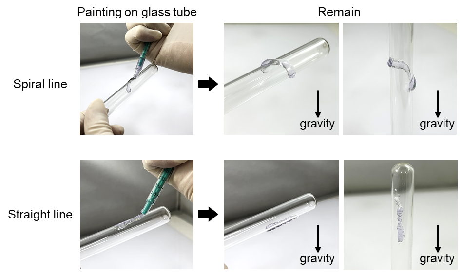
图S3 Photographs of the gelatin/dex-ald hydrogels applied in a spiral or straight line on a glass tube.
2.2. 明胶/地塞米松水凝胶的粘附性
作者的目标是开发一种粘附性水凝胶心脏补片,它不需要手术缝合,并且稳定地支持心外膜的机械性能。因此,他们研究了明胶/Dex-ald水凝胶的粘附性。将由10%明胶和5%地塞米松组成的水凝胶注射到一块组织块上,然后将另一块组织块覆盖到水凝胶上(图3A)。该凝胶对猪心外膜和皮肤具有较强的界面粘附力,分别为4.2±0.3kPa和19.5±1.7kPa(图3A和图3B)。不同相对分子质量(150 kDa、500 kDa和1.5 mda)的地塞米松制备的水凝胶之间的粘接强度没有显著差异(图S5)。值得注意的是,他们的可绘制水凝胶的组织粘附性与其他粘附性心脏补片相当。此外,水凝胶粘合组织的循环伸长没有引起粘合强度的实质性变化(图3C),显示出优异的耐久组织粘附性。经50次拉伸后,明胶/地塞米松水凝胶对心外膜和皮肤组织的初始搭接剪应力分别为96.5%和88.4%。在扭转、弯曲和水冲洗等各种变形下,水凝胶仍然牢固地附着在心外膜上(图3D)。总之,作者研发的明胶/地塞米松水凝胶有望稳定地附着在心外膜上,稳定地保持其形状,并适当地发挥无缝线心脏补片的功能。
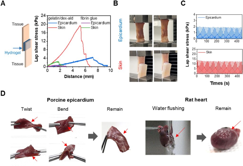
图3 Adhesion properties of the paintable hydrogels. (A) Lap shear stress of hydrogels to epicardium and skin tissues. (B) Photographs (distance = 0 mm and distance = 4 mm) of gelatin/dex-ald-tissue samples during the lap shear tests. (C) Cyclic stretching of gelatin/dex-ald-bonded epicardium and skin. Sample-bonded tissues were stretched up to 50 cycles with 0–4 mm of distance and 60 mm/min tensile rate. (D) Adhesion of (left) hydrogel-bonded porcine epicardium under dynamic conduction and (right) hydrogel-bonded rat heart with water flushing. Arrows indicate the hydrogels remaining on the tissue. Statistical significance was analyzed by one-way ANOVA followed by a Tukey analysis.
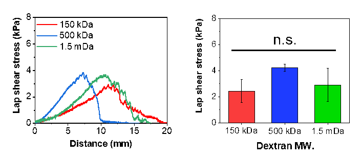
图S5 Lap shear stress of various hydrogels prepared with 5% gelatin and 5% dex-ald prepared with different MWs of dextran (150 kDa, 500 kDa, and 1.5 MDa) using epicardium tissue.
可涂和黏附的水凝胶心脏补片是一种创新的医疗器械,通常用于心脏手术中。这种补片具有特殊的水凝胶材料,可在手术中轻松涂覆和黏附于心脏表面。不断实验,不断迭代,用多样化的记录,挑战细微差别,然后通过耐心实现突破,这几点满足所有器械的创新。创新是引领行业高质量发展的第一动力。2023上海医疗器械创新展Medtec创新展全方位触达研发难点!特设“医工结合-数字医疗器械创新发展论坛”、“有源医疗器械研发与标准、质控论坛”、“创新材料论坛”等系列创新专题活动,还有专精特新企业带来具有行业或区域的独特性的技术与产品。立刻点击预登记,加入2023上海医疗器械创新展Medtec创新展。
2.3. ANGPTL4从明胶/地塞米松水凝胶中的释放
作者期望ANGPTL4具有高负载能力和的缓释能力,所以他们将A4/明胶/地塞米松水凝胶在酸性(pH 5.8)或中性(pH 7.4)缓冲溶液中孵育,分别代表了缺血和正常心脏组织。对照组可见ANGPTL4突发性释放。例如,初始负载的ANGPTL4在3 h内在pH 7.4中释放了76.7%。相反,A4/明胶/Dex-ald在早期孵育时间点表现出持续释放曲线,没有明显的突释(图4B)。ANGPTL4从第7天开始达到饱和释放,说明ANGPTL4在水凝胶中保持稳定。A4/明胶/Dex-ald在酸性缓冲液(pH 5.8)中释放ANGPL4的速度快于中性缓冲液(pH 7.4)。结果表明,在酸性条件下,ANGPTL4与水凝胶骨架之间的席夫碱会逐渐失稳,从而促进ANGPTL4的释放。由于心肌梗塞心脏表现出轻微的酸性,ANGPTL4的释放可能取决于心肌梗塞的严重程度(图4C)。此外,明胶/Dex-ald的模数逐渐降低,水凝胶在酸性溶液中逐渐降解,证实了Schiff碱在酸性微环境中的不稳定性(图S6)。接下来,我们在随后的实验中评估了体内降解情况。
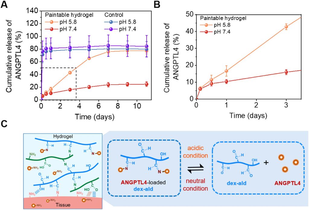
图4 In vitro release of ANGPTL4 from A4/gelatin/dex-ald hydrogels and gelatin hydrogels. (A) Cumulative release profiles of ANGPTL4 from the gelatin/dex-ald and gelatin control (crosslinked with glutaraldehyde) during the incubation. In vitro release tests were performed using Cy5-tagged ANGPTL4 in release buffer solutions (pH 5.8 and 7.4). (B) Inset of (A). (C) Schematic illustration for sustained release of ANGPTL4 through reversible Schiff base between dex-ald and ANGPTL4.
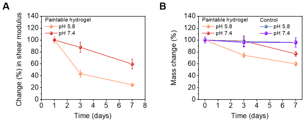
图S6 Stability of paintable hydrogels (gelatin/dex-ald) at different pH buffer solutions. (A) Change (%) in shear modulus (G’) of paintable hydrogels in release buffer solutions (pH 5.8 or 7.4). (B) Mass change (%) of paintable hydrogels and control hydrogels (glutaraldehyde-crosslinked gelatin) in release buffer solutions (pH 5.8 or 7.4).
2.4. 明胶/地塞米松水凝胶体外培养细胞
乳酸脱氢酶检测显示,在明胶/地塞米松水凝胶中培养的CMS显示出高达7天的良好细胞活力(图5A)。此外,在Transwell插入物中以水凝胶作为间接接触模式培养的CMS具有高达7天的高存活率(>95%)(图5B)。活/死染色表明,在带有水凝胶的Transwell插入物中培养的CMS与在组织培养板上培养的CMS之间的细胞活力没有显著差异(图5C和5D)。进一步研究了原位形成的明胶/地塞米松的细胞相容性,将水凝胶(混合前驱体溶液后5分钟) 放置到在磷酸三钙中培养的CMS上(图S7)。孵育48 h后,细胞活力与对照组(不加水凝胶的胞质干细胞)无显著差异(图S7),表明明胶和地塞米松没有潜在的细胞毒性。如图5E所示,在水凝胶上培养的CMS在培养的第7天观察到α-肌动蛋白的高表达和明显的肌。结果表明,他们研发的明胶/Dex-ald水凝胶具有良好的细胞相容性,并支持CM成熟。
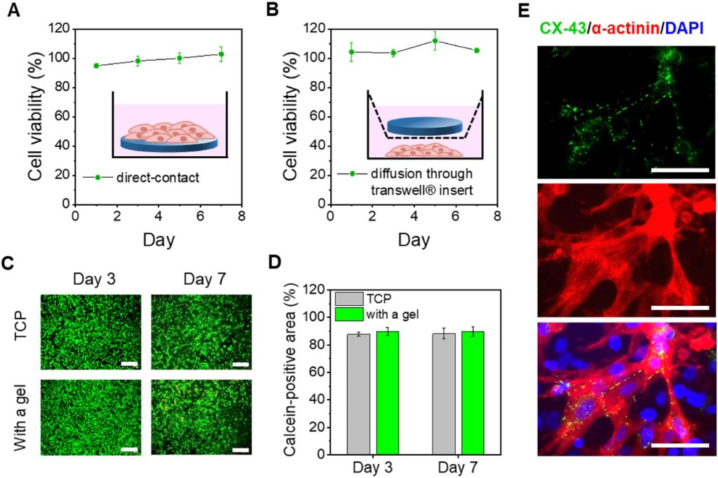
图5 In vitro cytotoxicity tests. LDH assay of (A) CMs directly cultured on the gelatin/dex-ald hydrogel and (B) CMs cultured in the transwell insert with the gelatin/dex-ald hydrogel (indirect contact). (C) Live (green)/dead (red) staining images of the CMs cultured on TCP or in the transwell insert with the hydrogel for 3 and 7 days. Scale bars = 200 μm. (D) Calcein AM positive area per image in each group. (E) Immunofluorescence staining for α-actinin (red), CX-43 proteins (green), and nuclei (blue) of the CMs cultured on the gelatin/dex-ald hydrogel for 7 days. Scale bars = 50 μm.
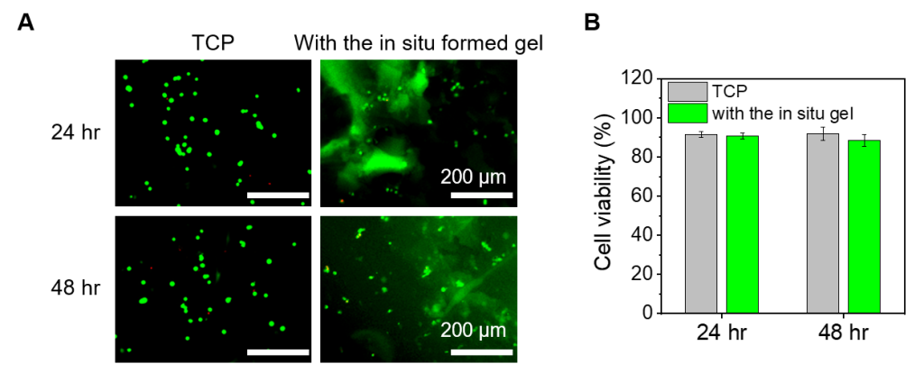
图S7 Cytocompatibility of paintable hydrogels (gelatin/dex-ald). (A) Live (green)/dead (red) staining images of the CMs cultured on a tissue culture plate (TCP) with or without coverage of the in situ formed paintable hydrogels. Fluorescence images were acquired 24 and 48 h after coverage. (B) CMs viability (%) in each group. For the cytocompatibility studies with in situ formed paintable hydrogels, the CMs were seeded on TCP (2.0 × 10⁵ cells) in 6 well culture plates and incubated for 24 h in CM growth medium. Then, a precursor solution of gelatin/dex-ald hydrogel was mixed and incubated in the petri dish at 37°C for 5 min. The in situ formed hydrogel was then carefully placed on the CMs growing on TCP. After addition 24 and 48 h incubation, the hydrogels covering the CMs were carefully removed, and the CMs were stained by Live/dead staining.
2.5. 明胶/地塞米松的体内着色性和降解性
作者使用双注射器将地塞米松(5%)和明胶(10%)溶液装入单独的桶中并注入混合。绘制的水凝胶成功地覆盖了整个左室,包括梗死区,并在心脏跳动的左室保持完好(图6A)。通过在治疗后7天和14天(图6B)采集心脏组织来检测所绘制的明胶/地塞米松水凝胶的体内生物降解性,结果表明所绘制的水凝胶逐渐降解。在第7天和第14天,心脏上最初的补片分别保留了大约46.6%和30.0%,这表明所绘制的明胶/地塞米松补片在体内可降解。总之,作者证明了明胶/地塞米松在心脏补片应用中具有适当的可涂性和体内降解性。
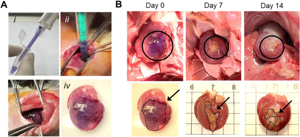
图6 In vivo studies for paintability and degradability of gelatin/dex-ald hydrogels on the heart. (A) Photographs of the procedures for painting the gelatin/dex-ald hydrogel as an epicardial patch; (i) mixing of gel precursor solutions using a dual syringe, (ii) transfer of the mixed hydrogel onto a rat epicardium, (iii) painting of the hydrogel on the rat epicardium, (iv) the painted hydrogel cardiac patch on the heart. (B) Photographs of the hearts painted with gelatin/dex-ald hydrogels on day 0, day 7, and day 14. Arrows indicate the hydrogels remaining on the hearts.
2.6. ANGPTL4可涂抹水凝胶用于心脏修复
为了确定A4/明胶/地塞米松贴片是否改善了心肌梗死后的心功能,作者在心肌梗死后第14天用超声心动图测量了心室收缩功能。如图7A所示。有代表性的超声心动图显示MI组左室内径增大,室间隔和后壁运动减少,明胶/地塞米松和A4/明胶/地塞米松组的室间隔和后壁运动减弱(图7B)。数据表明,假手术组和心肌梗死组的EF(%)分别为61.4±2.5和22.7±1.8,而明胶/地塞米松和A4/明胶/地塞米松组的EF(%)分别为31.6±2.9和36.7±2.8(图7C)。FS(%)值显示出与EF(%)相似的趋势(图7D)。用马森三色染色(图7E)分析MI诱导的纤维性瘢痕的形成。A4/明胶/地塞米松治疗组肝纤维化面积显著小于MI组和明胶/地塞米松组(图7F)。此外,补片治疗组左室壁厚度的减少有所减轻,其中A4负荷的心脏补片更有效(图7G)。这些结果表明,水凝胶贴片治疗和ANGPTL4局部给药通过为梗死心脏提供机械支持和消炎治疗,改善了心肌梗死后的心脏重构和功能障碍。
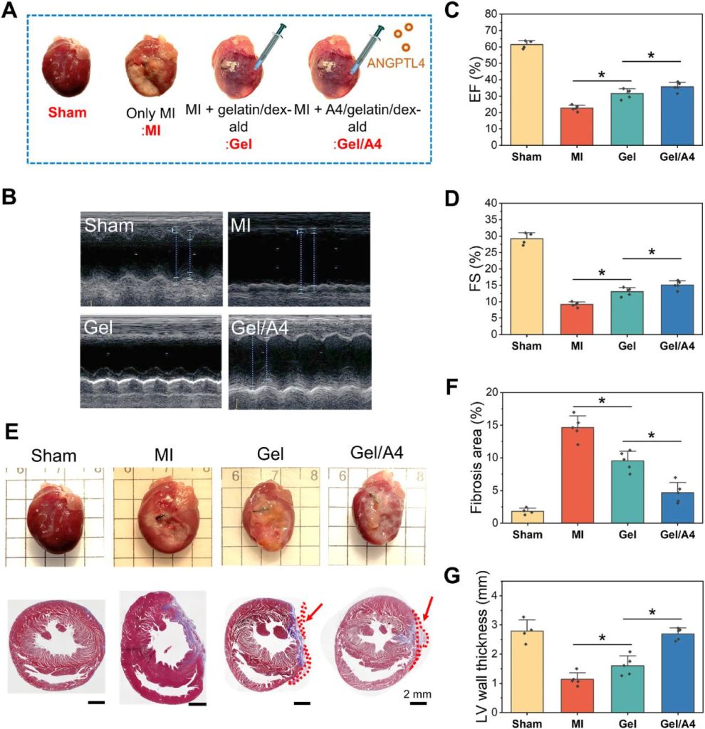
图7 Effects of gelatin/dex-ald hydrogel cardiac patches on heart functions and remodeling after MI. (A) Photographs of experimental groups (sham, MI, gelatin/dex-ald, and A4/gelatin/dex-ald). (B) Representative echocardiographic images after 2 weeks MI and treatment. (C, D) The LV function parameters (EF and FS) in various groups after 2 weeks MI. (E) Representative heart photographs and Masson’s trichrome staining images of the isolated hearts after 2 weeks MI and treatment. Arrows indicate the hydrogels remaining in the heart. (F) Fibrosis area and (F) LV wall thickness in each group. n = 4 (sham), n = 5 (MI), n = 5 (gelatin/dex-ald), and n = 5 (A4/gelatin/dex-ald). Statistical significance was analyzed by one-way ANOVA followed by a Tukey analysis. *p < 0.05.
接下来,作者使用α-SMA和vWF免疫荧光染色来确定水凝胶贴片在血管生成中的作用。与MI组和明胶/地塞德贴片组相比,A4/明胶/地塞尔德贴片治疗显著增加了新生血管的形成,特别是在边界区域(图8A)。此外,通过CD68和CD206染色来评估巨噬细胞的类型和分布(图8B)。A4/明胶/地塞米松治疗组心肌梗死区抗炎巨噬细胞多于明胶/地塞米松治疗组,提示ANGPTL4水凝胶补片具有抗炎作用。总体而言,与明胶/地塞米松组相比,ANGPTL4水凝胶贴片显著改善了心功能,抑制了心肌纤维化,促进了血管生成,并使巨噬细胞极化为抗炎活性。
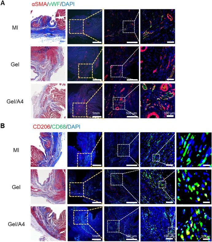
图8 Representative immunofluorescence images of rat heart tissues 2 weeks after MI. (A) Immunofluorescence staining of vWF (an endothelial marker, green) and α-SMA (a vascular protein marker, red) in the border zone. (B) Immunofluorescence staining of CD68 (a macrophage marker, green) and CD206 (an anti-inflammatory macrophage marker, red) in the border zone. The yellow color in the fluorescence images indicates the co-localization of CD68 and CD206. The upper images are the corresponding images of Masson’s Trichrome staining in each group (A and B).
3. 结论
作者成功地开发了生物相容性和粘附性水凝胶,使其能够使用10%的明胶和5%的地塞米松通过希夫碱形成原位凝胶。这种水凝胶适用于心脏补片,这种补片可以通过稳定的组织粘连而不缝合地附着在心外膜上,为心肌梗死后有效的心脏修复提供组织样的柔软和机械支持。此外,组织再生因子ANGPTL4被负载到明胶/地塞米松水凝胶上,用于局部和持续给药。ANGPTL4水凝胶贴剂可促进梗死心脏功能恢复,防止病理性组织重塑。免疫组织化学分析表明,ANGPTL4水凝胶贴剂促进了梗塞心肌血管生成,增加了抗炎巨噬细胞的数量。他们研发的ANGPTL4粘附性水凝胶贴片对心脏修复后脑梗塞有好处,也可能为未来的技术提供有用的信息,这可能会导致心血管疾病再生疗法的更好发展。
其设计旨在提供可调整的黏附性能,以确保在手术过程中牢固粘附,同时不损害心脏组织。这项技术在心脏修复手术中发挥关键作用,为外科医生提供了一种便捷而有效的方法,以支持患者的心脏健康。医疗器械中的研发创新至关重要,因为它推动着医学科技的不断进步。创新使医疗器械能够更有效地诊断、治疗和监测疾病,提高患者的治疗效果和生存率。新技术的引入也有助于改善医疗程序的安全性、便捷性和成本效益,推动医疗体系的发展。2023上海医疗器械创新展Medtec创新展致力于推动并传播创新研发,只有通过不断研发创新,医疗器械行业才能够应对不断演变的医学挑战,为患者提供更先进、更个性化的医疗解决方案。
文章来源:BioactMater生物活性材料